 Co., Ltd.")
Research progress on the internal structure and surface modification of 3D printed porous tantalum in bone tissue engineering
Release time:
2025-09-01
In today's medical context, the treatment of bone defects remains an important clinical problem [1]. Large bone defects are usually unable to be repaired by the body's own self-repair ability [2]. Therefore, the key problem in the treatment of bone defects is the lack of fusion between the graft and the bone, and the lack of rapid and sufficient vascularization, resulting in slow or failed bone regeneration. Therefore, the research on bone grafts remains a focus of research in the field of tissue engineering [3] and has great application prospects. Compared with dense materials, porous structure materials have some advantages, such as adjustable density, strength and elastic modulus to match bone tissue. Porous material implants can enhance the osteogenic response at the bone defect site [4]. Among the many porous structural materials, porous tantalum has a high porosity and interconnected pore structure, with a pore size range of 300 to 600 μm and a porosity of 75% to 85% [5-6]. Compared with other bone grafts, porous tantalum is closer to natural cortical bone [7]. In addition, porous tantalum has high corrosion resistance and biocompatibility [8-9], which can promote the formation of new bone inside. Its low elastic modulus and high friction coefficient can effectively avoid the stress shielding effect [10-12], minimize marginal bone loss, and ensure the primary stability of bone reconstruction and shaping. In addition, the surface of porous tantalum has high wettability and surface energy, which can promote the adhesion, proliferation and mineralization of osteoblasts [13-14]. Tantalum is more excellent in mechanical properties than other commonly used medical metal materials [15]. As a porous structural material, porous tantalum has many advantages in the treatment of bone defects, including regulatory properties that match bone tissue, promoting osteogenic response, high biocompatibility and excellent mechanical properties. Therefore, porous tantalum has great potential in the research and application of bone grafts. 3D printing technology has the advantages of fast modeling speed, high precision and the ability to achieve personalized customization according to needs [16]. In the preparation of porous tantalum, 3D printing technology can achieve precise control of porous structures and the preparation of complex shapes by precisely controlling printing parameters and designing templates. In addition, 3D printing technology can also introduce other functional materials or bioactive substances during the printing process to further improve the performance and bioactivity of porous tantalum. Therefore, the development of 3D printing technology provides broad prospects for the preparation of porous tantalum. The purpose of this article is to review the surface modification methods and strategies that have been applied to 3D printed porous tantalum and analyze the current research results. In addition, this article will also explore the research progress of the internal structure and surface modification of 3D printed porous tantalum in bone tissue engineering.
1 Study on the biological properties and spatial structure of porous tantalum
1.1 Biological properties of porous tantalum
In clinical practice, trauma, bone tumor resection, hip or knee replacement revision, etc. often lead to bone defects. Osteoblasts and osteoclasts in bone tissue maintain bone homeostasis by balancing bone resorption and bone growth. These two cells are very sensitive to mechanical stimulation. Therefore, changing mechanical factors can regulate bone growth and resorption, thereby promoting postoperative bone healing [17]. The selected implant material should have appropriate mechanical properties and osteoinductive properties, so it is crucial to select a suitable implant material. Recent studies have found that metallic tantalum has the characteristics of bioinertness, low toxicity and high corrosion resistance [18], and is considered to be a metal material with great potential. However, the elastic modulus of dense tantalum metal implants is significantly higher than that of human bone tissue [19], which can lead to stress concentration and stress barrier problems, thereby causing osteolysis at the implant site and even loosening of artificial prostheses [20]. With the rapid development of 3D printing technology in recent years, researchers have begun to use this technology to manufacture 3D printed porous tantalum. 3D printed porous tantalum has an elastic modulus and porous structure similar to cancellous bone[21]. Compared with dense tantalum metal implants, the elastic modulus of 3D printed porous tantalum is closer to that of human bone tissue[22], thereby reducing the problems of stress concentration and stress barrier, and helping to improve the stability and long-term durability of the implant site. Wang et al.[7] explored the biocompatibility of porous tantalum. The researchers seeded bone marrow mesenchymal stem cells (BMSCs) onto porous tantalum scaffolds and incubated them in culture medium. Subsequently, they observed the porous tantalum scaffolds using a scanning electron microscope (SEM) at designated time points. Under high magnification, the number of cells increased on the 5th day compared to the results on the 3rd day of culture, and it was seen that the cells had grown into the interior of the porous tantalum and formed interconnected protrusions. This indicates that porous tantalum can promote the growth of bone cells into the implant, has osteoinductivity and biocompatibility, and can form a support structure to help bone tissue better integrate with the implant. In addition, Wang et al. [23] inoculated osteoblasts cultured in vitro onto porous tantalum. On the third day of culture, cells were observed to adhere to the surface and pore walls of the porous tantalum. On the seventh day, osteoblasts were observed to fuse into thin sheets, accompanied by cell-secreted matrix, which almost covered the surface of the porous tantalum. These experimental results show that porous tantalum not only provides space for cell adhesion and proliferation, but also promotes the secretion and penetration of cell metabolites, further proving that porous tantalum has good biocompatibility. Lu et al. [24] cultured bone marrow mesenchymal stem cells on porous tantalum. After 7 days of culture, under low magnification, it was observed that the cells formed a continuous layer on the surface of the porous tantalum and grew into the pores of the porous tantalum. Under high magnification, it was observed that these proliferating cells were irregular in shape, which further demonstrated that porous tantalum has non-toxicity and good biocompatibility. In addition, Dou et al. [25] found that porous tantalum can promote the adhesion and proliferation of bone marrow mesenchymal cells and promote the osteogenic differentiation of BMSCs in vitro. The study by Gee et al. [26] demonstrated that porous tantalum increased the proliferation of human fibroblasts in direct contact, and no quantifiable negative effects on human fibroblast behavior were observed within 28 days. This indicates that porous tantalum has no inhibitory effect on human osteoblasts or mesenchymal cells, and can even stimulate soft tissue healing at the tissue interface.
These research results support the application of porous tantalum in bone tissue engineering. Porous tantalum has good biocompatibility, can promote the adhesion, proliferation and secretion of bone cells, and integrates well with bone tissue. These findings lay the foundation for the application of porous tantalum as an implant material in bone defect repair and bone tissue engineering.
1.2 Spatial structure of porous tantalum
The physical properties of porous tantalum have been improved in commercial products, including high porosity (75% to 85%), dodecahedral pore structure and pore size of 400 to 600 μm. Reports show that numerous experimental results indicate that porous tantalum scaffolds with a pore size of 400 to 600 μm are more conducive to the ingrowth of bone tissue [27]. Scaffolds with an average pore size of 400 μm and a porosity of 70% can promote cell migration, proliferation, osteogenic differentiation, and the formation of blood vessels and bone tissue [28-29]. In terms of bone integration, the high pore size and porosity of porous tantalum contribute to the ingrowth of bone and soft tissue due to its extensive three-dimensional internal space and high pore interconnectivity. The high porosity of porous tantalum ensures the needs of vascularization and nutrient flow, thereby achieving early and rapid bone integration. In addition, porous tantalum has high wettability and surface energy, which can promote the adhesion, differentiation and diffusion of stem cells, osteoblasts, chondrocytes, vascularized fibrous tissue and tendons [30-31]. These properties indicate that porous tantalum has good biocompatibility and osteoinductivity, and has a positive effect on tissues such as bone and tendons. The physical properties of porous tantalum, such as pore size and porosity, have a significant impact on the success of bone tissue engineering. Selecting an appropriate pore size range is crucial for bone ingrowth and sustained penetration; pore size and porosity should be selected and optimized based on specific clinical needs and application objectives.
2 Surface Modification of Porous Tantalum
The inertness and low bioactivity of porous tantalum are a major challenge to its development in bone tissue engineering. To overcome these challenges, researchers have introduced various methods to modify the surface of porous tantalum to enhance its bioactivity and bone tissue integration, thereby promoting its further application in clinical applications. These methods can be primarily categorized into two categories: biomaterial coating and surface treatment.
Biomaterial coating is a commonly used surface modification method that modifies the surface properties of porous tantalum by applying additional layers to the surface. These coatings can include bioactive substances, drugs, cytokines, etc. to improve cell attachment, enhance osteoinduction, or inhibit bacterial infection. Common coating materials include hydroxyapatite (HAP), calcium phosphate tribasic (CAP), poly-ε-caprolactone (PCL), and polylactic acid (PLA). These coatings can improve the biocompatibility, bioactivity, and bone tissue integration of porous tantalum, thereby promoting bone defect repair.
Surface treatment is another commonly used surface modification method, which modifies the surface of porous tantalum through physical or chemical means. Common surface treatment methods include anodization, acid washing, solvent treatment, and plasma treatment. These methods can modify the surface morphology, roughness, pore size, and surface energy of porous tantalum, thereby enhancing its interaction with surrounding tissues and cells. Surface treatment can enhance cell attachment, osteogenic capacity, and bone tissue integration of porous tantalum.
These biomaterial coatings and surface treatment methods provide a variety of options for modifying porous tantalum to meet diverse clinical needs and application scenarios. However, the selection and optimization of each method still requires in-depth research and evaluation to ensure that the modified porous tantalum material has good biocompatibility, bioactivity and long-term stability in clinical applications. 2.1 Nanotube modification of porous tantalum surface In the study of Zhang et al. [32], they used electrochemical anodization to construct a unique nanostructure on the surface of porous tantalum. Under optical view, there is no obvious difference between ordinary porous tantalum and porous tantalum modified by nanotubes, both of which show regular and orderly trabecular structures. However, by magnifying the surface of the porous tantalum modified by nanotubes, a micro-rough morphology can be observed, and the protrusions on the surface are uneven in size. This nanotube-modified porous tantalum surface morphology can increase its interaction with surrounding tissues, cells and proteins. With a larger surface area and irregular surface morphology, it can provide more contact points and structural features, which helps to enhance cell attachment and growth, as well as protein adsorption and interaction. This is crucial for the successful integration and biocompatibility of porous tantalum in bone tissue engineering and implant applications. However, the long-term effects and biocompatibility of nanotube-modified porous tantalum require further research and evaluation. In addition, optimizing parameters such as nanotube size, distribution, and density are also important factors to consider in order to achieve optimal bioactivity and tissue compatibility.
2.2 Porous tantalum loaded with magnesium ions
Mg, as an essential element in the process of bone growth and development, has the ability to effectively promote osteogenesis and angiogenesis. In the study of Ma et al. [33], different concentrations of magnesium (Mg) were doped onto the surface of 3D-printed porous tantalum using the surface adhesion ability of polydopamine (PDA) to improve the surface bioactivity of porous tantalum. The study verified the effect of Ta-PDA-Mg scaffold through a series of in vitro and in vivo experiments. The experimental results showed that Ta-PDA Mg2 significantly enhanced the formation of vascularized bone and bone integration.
This method provides new directions and opportunities for the application of porous tantalum in bone defect repair and bone tissue engineering. However, the long-term effects and biocompatibility of magnesium-doped porous tantalum require further research and evaluation. In addition, optimizing the doping concentration and preparation process are also key factors that need to be considered to ensure the reliability and effectiveness of porous tantalum-Mg materials in clinical applications.
2.3 Surface coating modification of porous tantalum
Hydroxyapatite (HA) is a commonly used bone tissue supplement and filler, which is widely used because of its biocompatibility, biodegradability and non-toxicity [31]. In porous tantalum modification, calcium phosphate and HA have also been used for surface modification and drug delivery. Studies have confirmed that the use of alendronate-CaP coating to repair the gap between porous tantalum bone and implant can successfully fill the gap of simulated bone defects. In the presence of alendronate coating, the bioactivity of the porous tantalum implant surface is enhanced [34]. This successful repair mechanism is attributed to the slow release of alendronate, which inhibits osteoclast activity while enhancing osteoblast activity. Therefore, this method is considered to be an effective way to improve the osteoconductivity of porous tantalum [35].
In the biomedical field, polylactic acid (PLA) materials can be used as drug delivery materials, tissue engineering scaffold materials, bone repair materials, etc. It is currently a widely used synthetic polymer, and its manufacturing methods and properties have been widely studied [36]. PLA can increase bone conductivity and bone formation [37-38], and is biocompatible, biodegradable into non-toxic components, and its degradation rate is controllable after entering the human body [39]. In the study of Liu et al. [40], a new porous PLA/β-TCP/PDA/Ta scaffold was designed and manufactured. This scaffold has a reasonable physical structure, suitable mechanical properties, excellent bioactivity and the ability to promote cell proliferation, meeting the initial needs of bone regeneration and tissue repair. In the study by Zhou et al. [41], CaP nanospheres were mixed with PLA polymer to prepare a uniform suspension, and the surface of tantalum plates and porous tantalum scaffolds was modified. CaP-PLA containing vascular endothelial growth factor (VEGF) and transforming growth factor beta (TGF) was loaded on the porous tantalum scaffold. It was found that this composite scaffold can provide growth factors, physical support, and structural guidance for the growth of new bone, and is conducive to guiding subchondral bone regeneration. The application of the above coatings and materials has expanded the functionality and performance range of porous tantalum. By combining these materials with porous tantalum, its biocompatibility, corrosion resistance, oxidation resistance, and osteoconductivity can be improved, thereby further promoting bone defect repair. However, more research and experiments are needed to evaluate the effectiveness and long-term effects of these modification methods to ensure their safety and feasibility.2.4 Surface modification of porous tantalum for antibacterial properties Implant-related infection has long been a thorny problem in the clinical setting [42], which will lead to surgical failure and additional surgical expenses. Therefore, it is imperative to find a reasonable method to give porous tantalum antibacterial properties. Polyhydroxyalkanoates (PHA) are biodegradable and biocompatible materials that are widely used in drug delivery and tissue engineering [43]. In the study of Rodríguez-Contreras et al. [44], a polyhydroxyalkanoate (PHA) coating containing antibiotics was loaded onto the surface of porous tantalum to achieve controlled antibiotic release. This coating can avoid infection of porous tantalum implants and protect them from infection by Gram-positive and Gram-negative bacteria. In addition, Liao et al. [45] proposed a ZnO nanorod-nanosheet layered coating for porous tantalum surfaces. This coating has a two-stage release mode and can continuously release antibacterial substances in the body, thereby preventing implant-related infections. The design of the ZnO nanorod-nanosheet coating can prolong the duration of the antibacterial effect, especially in the critical period after surgery. In addition, Guo et al. [46] successfully inhibited the proliferation of chondrosarcoma cells cultured in vitro by integrating doxorubicin into the negatively charged trimer on the surface of porous tantalum and reacting it with positively charged methylated collagen. This method utilizes the interaction between the negative charge of the drug and the positive charge on the surface of porous tantalum to achieve an anti-proliferative effect and is expected to be used to prevent and treat tumor-related implant infections. In addition, a comparative study was conducted by Griseti et al. [47], who compared porous tantalum loaded with vancomycin with bone cement loaded with antibiotics and found that porous tantalum can prolong the release time of vancomycin in the body, increase the local antibiotic concentration level, and reduce the risk of infection around the implant. This approach avoids the systemic toxicity that may result from systemic antibiotic administration [48] and reduces the overuse of antibiotics, the development of multidrug-resistant microorganisms, and other unknown risks.
These studies demonstrate strategies for biological modification of porous tantalum surfaces to enhance their antibacterial properties. By integrating antibacterial substances or drugs into the porous tantalum surface, implant-related infections can be effectively prevented and controlled, surgical success rates can be improved, and additional expenses can be reduced. However, these methods are still in the research stage and further experimental and clinical studies are needed to verify their effectiveness and feasibility.
2.5 Biological modification of porous tantalum surfaces
In a study by Zhao et al. [10], a 3D-printed porous tantalum-gelatin nanoparticle-hydrogel composite scaffold was developed. This composite scaffold has biocompatibility and biomechanical properties and can promote the formation of highly vascularized bone tissue. The design of this composite scaffold combines the excellent mechanical properties of porous tantalum and the bioactivity of gelatin nanoparticles. A scaffold with complex structure and pores is manufactured by 3D printing technology, providing an environment required for bone cells to grow and vascularize. In the study by Wei et al. [49], they constructed an integrated biofabrication platform using mesenchymal stem cells, porous tantalum and chondrocyte/collagen membrane (CM) to repair osteochondral defects in the weight-bearing area of goats. The results showed that the chondrocyte collagen membrane maintained the characteristics of chondrocytes and highly expressed cartilage-related genes. This composite porous tantalum scaffold material showed good results in repairing bone defects. In the study by Wang et al. [50], RGD peptide (Arg-Gly-Asp Peptides, RGD) was modified on the surface of porous tantalum and used to repair segmental bone defects in rabbit radius. Compared with the unmodified porous tantalum scaffold, the RGD peptide-modified porous tantalum scaffold had increased bone formation at the interface and within the pores, and the biomechanical properties of the RGD peptide-modified porous tantalum group were better than those of the unmodified porous tantalum group. These studies have demonstrated the potential of biological modification of porous tantalum to promote bone defect repair. By combining porous tantalum with other biomaterials, it can offer improved biocompatibility, bioactivity, and mechanical properties, thereby promoting bone tissue regeneration and repair. These studies have broadened the application possibilities of porous tantalum and provided valuable insights for the development of more effective bone defect repair methods.
3. Deficiencies and Future Prospects
This article provides a detailed description of the characteristics and applications of porous tantalum as a bone defect repair material. Porous tantalum exhibits excellent biocompatibility and bone-like biomechanical properties, enabling effective bone growth at bone defect sites. Its high corrosion resistance, low toxicity, low elastic modulus, and high surface friction coefficient make it an ideal metal material for bone defect repair. A review of the literature reveals that porous tantalum exhibits enhanced osteogenic and angiogenic differentiation potential, promoting bone growth and osteoinduction. Consequently, porous tantalum has been widely used in bone tissue engineering, achieving satisfactory clinical results. However, further improvements in its performance are still needed.
With the advancement of 3D printing technology, 3D-printed porous tantalum offers new possibilities for the design and fabrication of novel porous tantalum-based implants for personalized medicine. By tailoring the macrostructure, pore size, pore geometry, and porosity of porous tantalum implants, patients' needs can be met. 3D-printed porous tantalum exhibits exceptional flexibility, particularly for large and complex bone defects in load-bearing areas. It outperforms other porous scaffolds in compression and deformation resistance, and possesses biomechanical properties approaching those of bone scaffolds. Although relatively little research has been conducted on 3D-printed porous tantalum, it holds broad promise for clinical orthopedic applications. Furthermore, modification methods can enhance the bioactivity and antimicrobial activity of porous tantalum. While surface modification techniques are continuously improving, most current research remains in animal experiments. Therefore, further experiments are needed to validate the biological and mechanical properties of porous tantalum and further applications in diverse biological and mechanical environments to demonstrate its significant value in orthopedic clinical practice. Only then can porous tantalum be widely used in bone defect repair and its clinical potential be realized.
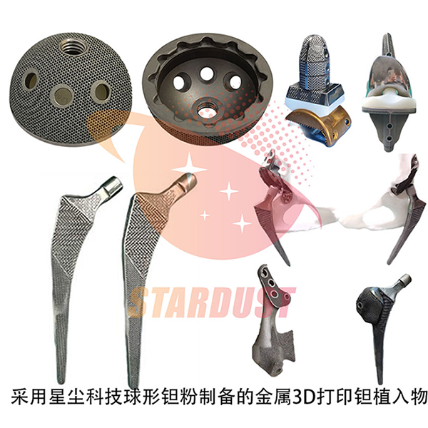
Medical-grade spherical tantalum powder
Stardust Technology uses radio frequency plasma spheroidization technology to produce spherical tantalum powder. With its excellent biocompatibility and bone ingrowth properties, it is widely used in 3D printing of clinical medical implants for spine, joints, and trauma. Stardust Technology pioneered the industrialization of medical-grade spherical tantalum powder in China. Working with leading domestic orthopedic hospitals and medical device companies, Stardust Technology has promoted the clinical application of tantalum metal. The company has participated in the development of national standards, including GB/T 38975-2020 Tantalum and Tantalum Alloy Powders for Additive Manufacturing, GB/T 41883-2022 Powder Bed Fusion Additive Manufacturing of Tantalum and Composite Alloys, YY/T1851-2022, an industry standard for medical pure tantalum powder for additive manufacturing, T/CAMDI 065-2021 Additive Manufacturing of Tantalum Knee Prostheses, and T/CAMDI 066-2021 Additive Manufacturing of Tantalum Personalized Bone Defect Fillers. Stardust Technology has assisted in the clinical application of tantalum metal in over 500 cases, including implants for tantalum intervertebral fusion devices, bone spacers, hip, shoulder, knee, and ankle joints.
http://en.stardusttech.cn/products/48.html
For more details, please contact Vicky Zhang at +86-13318326185

News
Stardust Technology (Guangdong) Co., Ltd.

101, Building 1, Liandong Youzhi Zone, Senshuji Road, Nansha Community, Danzao Town, Nanhai District, Foshan City,Guangdong Pro.,China
Tel:13318326187
13378621675
QR code